Jemel, B., Oades, R. D., Oknina, L., Achenbach, C. & Röpcke, B., Frontal and temporal lobe sources for a marker of controlled auditory attention: the negative difference (Nd) event-related potential . Brain Topography, 15, (4) 249-262 . - (request a pdf copy).

In accord with our understanding of journal policy, we present the pre-publication text (view).
The original publication (doi:10.1023/A:1023915730566) is available at http://springerlink.metapress.com/content/lxhnwx7bkma2/?p=c87874c9b69845fda1d9cd9ca76adfb6&pi=21

Introduction:
In this study we sought frontal and temporal lobe dipole sources associated with event-related potential (ERP) marker of auditory controlled attention processes, known as the Negative Difference (Nd) -- for comparison with activity arising earlier in association with the detection of stimulus change (Mismatch Negativity, MMN, Jemel et al., 2002).

(We have previously reported on a) the normal topography of Nd (Oades et al., 1995) and its development over childhood and adolescence (Oades et al., 1997), and b) on an asymmetric Nd topography in paranoid and non-paranoid subgroups of patients with schizophrenia. While the differences can appear to indicate a reduced amplitude (Oades et al., 1994), they may reflect changes of the latency of regional activation (Oades et al., 1996).

Methods:
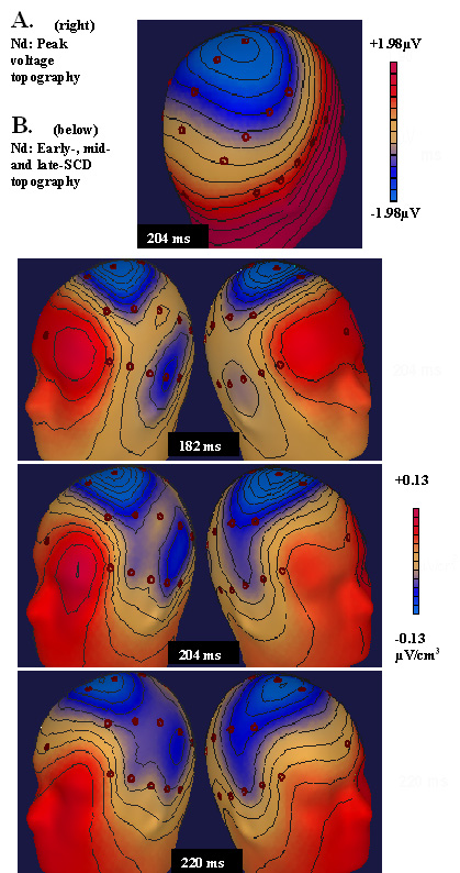
In two sessions a month apart (T1 and T2), 14 healthy subjects were presented with a 3-tone oddball passively, then as a discrimination task. EEG recordings were taken from 32 sites. Nd was calculated by subtraction of the ERP elicited by a non-attended deviant stimulus (session 1) from that after the same frequency-deviant tone as a target in session 2. Putative generators in the 180-228 ms post-stimulus latency rangesFigure 1 were modelled with brain electrical source analysis (BESA) and mapped to the modified Montreal brain-atlas (Garneron algorhythm).

Fig. 1 SCD maps at 3 time points in the latency window. .

Results:
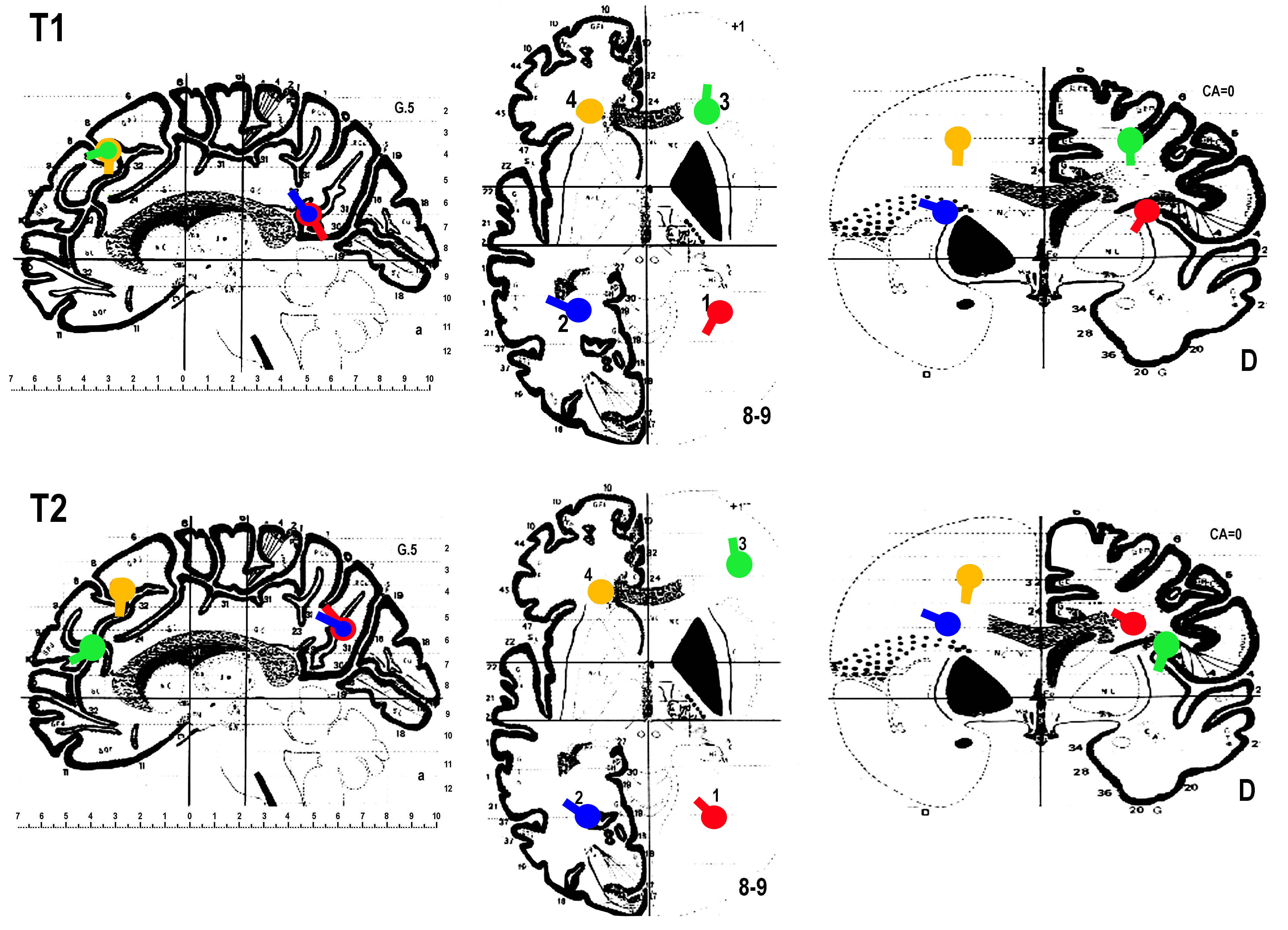
First:
Initial T1 analyses located Nd dipoles bilaterally in the superior temporal gyrus (BA 22), and the dorso-lateral prefrontal cortex (BA 8). Figure 2
Second: Re-test in T2 allowed estimates of the temporal and spatial extension of activity. (Peak activity occurred 14 ms later: T1 and T2 dipole strengths were well-correlated.). Step-by-step analysis showed the best spatial fit for the inverse-solutions extended 3-6 mm from the modelled point sources, - but for temporal lobe sources this increased 15 mm caudally.
Third: The sources lay a) in the right mid-frontal cortex (BA 10), that was rostral and ventral to that in b) the left superior frontal gyrus (BA 8). c) The two so-called temporal lobe sources extended over the temporo-parietal-occipital border (approx. BA 39). (see Fig. 2 on the right, here)

Conclusions:
Nd measures of controlled selective attention were localised in regions of the brain that imaging studies have associated with sustained attention, problem-solving and working memory.

Frontal Nd sources were more dorsal on the right and more rostral on the left than MMN dipoles (automatic change detection) reported for the right inferior frontal and left anterior cingulate regions. Temporal lobe sources were active later than MMN and frontal Nd dipoles, and lay more posterior and medial than MMN dipoles.

We propose the following potential sequence of activation (for auditory tones) after the initial N1 excitatory response: For noting the "changed stimulus" - an MMN is initiated in the superior temporal gyrus (perceptual template for standards), followed by deviant registration information passed to the right inferior frontal (for a potential switch) and left cingulate (conflict/priority resolution). For noting the "changed relevance" Nd is initiated in the right medial frontal region (close to the MMN dipole), and the information is passed to left superior frontal regions for planning response, and posterior temporal comparator processes.